Western Blot is a classic technique in Biological science research, yet many researchers frequently encounter various issues during the experiment, such as blurry bands, high background noise, or signal loss, leading to suboptimal results.
This article provides a detailed analysis of the ten most common problems in Western Blot experiments and offers practical solutions to help you overcome these challenges and ensure clear and precise results!
Problem 1: No Background Signal or Bands
Possible Causes:
The primary antibody may have lost its activity, or the secondary antibody may be incompatible with the species, such as the rat (Ra) antibody was replaced with a rabbit (Rb) antibody.
Solutions:
- 1.Check whether the sample membrane is within the imaging range.
- 2.If the target protein shows no signal while the Internal Control Protein is normal, the primary antibody may have lost its activity, or the wrong secondary antibody was used. Try re-incubating with the correct antibody.
- 3.If neither the target protein nor the internal control protein shows a signal, consider whether the chemiluminescent substrate has expired. Prepare fresh chemiluminescent substrate; if bands still do not appear, repeat the experiment, the transfer may have failed.
- 4.If faint bands are visible, the protein loading amount may be too low, or the primary antibody concentration is insufficient.
Problem 2: High Background signal
Possible Causes:
Insufficient blocking, excessively high primary antibody concentration, or inadequate washing time and frequency.
Solutions:
- 1.Reduce the primary antibody concentration to 1:5,000 or 1:10,000.
- 2.Increase the washing time and frequency (e.g., 5 minutes × 5 times or 10 minutes × 3 times).
- 3.If the issue persists, re-block the membrane.
Problem 3: Heterogeneous Background signal
Possible Causes:
The PVDF or NC membrane may not have been kept consistently moist during the experiment, or the temperature may have been too high during electrophoresis or transfer.
Solutions:
- 1.Ensure that the protein side of the membrane does not dry out during the experiment.
- 2.Avoid using excessively high voltage during electrophoresis and transfer, as it can cause elevated temperatures.
- 3.When handling the membrane, use tweezers to grip the area where the marker is located to prevent damage to the protein sample.
Problem 4: Black Dots and Streaks
Possible Cause:
The blocking solution may not have fully dissolved, leading to insoluble particles.
Solutions:
- 1.When preparing the blocking solution, ensure the non-fat dry milk is thoroughly dissolved by shaking well.
- 2.After blocking, wash the membrane with PBST 1-3 times.
Problem 5: Non-specific protein binding bands
Possible Cause:
The primary antibody may bind non-specifically to proteins, which is more common with polyclonal antibodies.
Solutions:
- 1.Extend the blocking time.
- 2.Replace the primary antibody or reduce its concentration.
Problem 6: White Circles in the Bands
Possible Cause:
Bubbles in the PAGE gel or between the membrane and gel during the transfer process.
Solutions:
- 1.When preparing the gel, slowly and evenly add the lower gel using a syringe.
- 2.Remove any bubbles between the membrane and gel before the transfer.
- 3.The entire transfer sandwich (including filter paper, gel, and membrane) should be assembled in transfer buffer to ensure that all components remain moist.
- 4.Use a roller to remove bubbles.
Problem 7: White Area in the Middle of Bands
Possible Cause:
High enzyme concentration in the center may deplete the substrate too quickly, causing the center to lose luminescence after substrate consumption.
Solutions:
- 1.Use rapid exposure to capture the image before the substrate in the center is consumed.
- 2.Reduce the protein amount and decrease the concentrations of the primary and secondary antibodies.
Problem 8: Band Smearing
Possible Cause:
Excessive protein amount or slight degradation, high primary antibody concentration, or overly long incubation time.
Solutions:
- 1.Adjust the protein amount.
- 2.Reduce the primary antibody concentration and incubation time.
Problem 9: Dumbbell-Shaped Bands
Possible Cause:
Uneven polymerization of the gel or sample impurities.
Solutions:
- 1.Wait for the PAGE gel to completely polymerize before removing the comb.
- 2.Centrifuge the sample before use to avoid impurities settling in the center.
Problem 10: Distorted Bands
Possible Cause:
Bubbles or insoluble particles present in the SDS-PAGE gel.
Solutions:
- 1.Be cautious during gel preparation and use impurity-free liquids.
- 2.Replace the sponge pad used for gel preparation.
- 3.When pouring the gel, use a gel pipette or syringe to add the gel, and if bubbles form, remove them with a syringe.
Most common issues in Western Blot experiments can be resolved by optimizing experimental conditions and techniques.
Thanks for reading! If you encounter other problems during your experiment, feel free to leave a comment, and we will provide professional assistance!
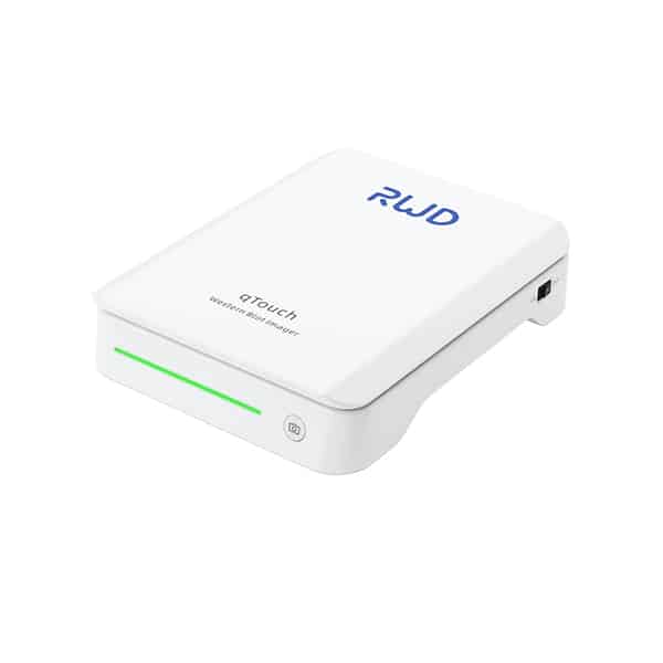
RWD Western Blot Imager
GET FREE DEMO for RWD Western Blot Imager
- 1.Ultra-high Sensitivity: Suitable for pg-level samples, providing clearer imaging of weak signals. Phosphorylated proteins can also be perfectly visualized.
- 2.Ultra-fast Imaging: Most samples can be imaged in just one second, improving experimental efficiency.
- 3.Wide Imaging Range: Both strong and weak signal bands can be developed simultaneously.
- 4.Compact Size: Saves laboratory space with its small design.
- 5.Greater than 150 cm² Photosensitive Chip: Contact imaging with no signal loss.
- 6.Intelligent Software: Built-in collection and analysis features, with direct visibility of protein band grayscale values.